My mother’s pathology – sarcoidosis

A lifetime of medication, and side-effects. At some point my mother began to store package inserts of the drugs she was prescribed, annotated with her reactions to them.

At 30 years old my mother had a radical mastectomy, meaning her left breast was removed along with the chest wall muscle underneath, and the lymph nodes under her left arm. Years later as a medical student I learned that she had not needed this extreme surgery. But back then the distinctions between types and stages of breast cancer were still being worked out, and my mother and her surgeon opted not to take any chances.

The mutilating operation changed the course of her life. She also underwent some form of ovarian ablation (her ovaries were rendered non-functional so that they produced no hormones which might stimulate the cancer to grow.) This brought on early menopause, further adding to the negative effects on her physical health and psyche.

Ten years after the breast surgery she complained of chest pain and a cough. Her chest X-ray showed a “white-out”, a blizzard obliterating both of her lungs. As I remember it, the possibilities were lymphangitis carcinomatosa (indicating cancer finely spread throughout the lungs), bird fancier’s lung (an allergic reaction to her parrot), tuberculosis (a chronic bacterial infection) or sarcoidosis (an inflammatory disease with no known cause). She had a bronchoscopy (for which she refused anaesthesia or sedation) and the biopsy sample was compatible with TB or sarcoidosis, since they have a very similar appearance under the microscope. Because tuberculosis is rife in South Africa, she was started on a course of anti-tuberculous drugs, which made her very unwell. The polymerase chain reaction (PCR) was a brand new form of testing back then, but training as a virologist I was able to arrange a PCR to detect Mycobacteria tuberculosis in the biopsy tissue, which proved negative. Her treatment was changed to the only available option for sarcoidosis, which was high dose prednisone. She fought to have the dose reduced to a level where the side-effects were tolerable. After more than a decade of treatment the disease seemed to “burn out”, leaving her with lung fibrosis (scarring) and bronchiectasis (dilated airways).

There are many similarities with my mother’s story in this case of pulmonary sarcoidosis from our collection. However this patient died soon after diagnosis.

By now, in her 50s, her skeleton had become dangerously osteoporotic (brittle), this because of the long-term prednisone treatment and premature menopause. Attention turned to her bone density and a number of drugs were tried to improve it, but all of them induced problematic side effects.

In her late 60’s chest symptoms occurred again, and a right-sided pleural effusion (fluid between the lung and the chest wall) was found. This needed repeated drainage and finally the pleural space was simply eliminated by fusing the lung to the chest wall, using a chemical irritant. By now she also had signs of pulmonary hypertension and cor pulmonale (right heart failure), and required a pace-maker for atrial fibrillation (heart flutter).

At age 73 the final event was sudden. Overnight she became desperately ill, and in the emergency unit her oxygen saturation was low. Laboratory tests showed that her acute pneumonia was caused by parainfluenza-3 virus, a virus usually seen in very young children. She required high flow oxygen in intensive care and was given massive doses of cortisone to try to prevent further inflammation in her lungs. She sank into delirium with a steroid psychosis. Already weakened by pulmonary cachexia (wasting), she stopped eating and then drinking.

Cause of death on on her death certificate was “end-stage lung disease”. I don’t recall if sarcoidosis was specifically mentioned.

There is little lung with which to breathe in this case of long-standing pulmonary sarcoidosis. There is fibrosis (scarring) of the pleural surfaces as well as a lot of fibrosis around the bronchi (airways) within the lung. Fibrosis of blood vessels within the lungs causes pulmonary hypertension.

At most, 1 in 20 people diagnosed with sarcoidosis will die of the disease, usually of respiratory failure. Although the inflammatory response in sarcoidosis has been well studied, the actual cause of the disease remains unknown. Aside from the lungs, it commonly involves lymph nodes, skin and liver, and less often other organs such as the heart and eyes. 

The full story of any disease is really only known through personal experience of it.

The manner of our passing

In the time of COVID-19 you can be forgiven for thinking people are dying of one thing only. In a war, injury would seem the most likely way to die. Ancient infectious diseases still bring death today. Inhabiting a pathology museum you’d conclude that malignancies are what kill most people. At different times and in different places, different causes of death dominate in reality or in our imagination.

Does it matter what a person dies of, or just that they have died? Perhaps it should just be the fact of their death that is important, but I would argue that instinctively one wants to know why, or of what. Is it because the cause of death tells us something about a person’s dying, the story of their last days or hours? Perhaps it is because what others are dying of presages what will kill us.

On an individual level, we hear of a person’s death, but so often the cause is not shared. Announcements of death may give hints, such as ‘suddenly’, ‘peacefully’ or ‘after a long (or short) illness’, these differentiating a relatively few precipitous events from the many causes of slow decline. The word ‘tragically’ might flag death by accident or suicide. When the cause of death is clearly stated, this may be offered to accurately inform, or might be to avert speculation and potential stigma.

On a societal scale, accurate figures for cause of death can be difficult to source, since even official agencies have trouble collecting them. In South Africa the current top killers are HIV/AIDS, heart attacks, strokes, diabetes, tuberculosis, inter-personal violence and road accidents (1). There are of course a myriad other diseases and events which can be fatal, some of which you’d be exceptionally unlucky to succumb to. Our collection here in the Pathology Learning Centre is a reminder of such things.

Some very rare medical misfortunes, all fatal. Left: the tattered brain of SSPE, a late complication of measles. Centre:  Budd-Chiari syndrome  Right: a tumour carried in a transplanted kidney .

So most of us will die as dictated by statistical probability. Even during wars, combatants and civilians have mainly died of disease rather than violent injury (2). Inevitable lifestyle diseases claim most lives in developed nations. The number of cancers in a pathology museum reflects pathology’s diagnostic focus, rather than cancer’s frequency. Despite perceptions and media attention, COVID-19 accounts for a small quota of death in the 21st century so far and a vanishingly small fraction in human history (3).

People die every second of every day, and each of us will die – this matters to us, and almost certainly it matters to us what of.

Notes

  • 2. Ellis J. The Sharp End: The fighting man in world war II. New York: Scribner’s; 1980.

Of mushrooms and men

Of the many natural hazards in Africa, mushrooms are not necessarily the first thing to spring to mind. But Neil van der Reij, a mushroom researcher with the Department of Agriculture and Environmental Affairs, is quoted as saying mushroom poisoning is “common” in South Africa (1).

In our collection we have two liver specimens titled “mushroom poisoning”. Each case is noted to be “one of a whole family who were poisoned by eating mushrooms”. The incident occurred in Cape Town in May of 1927.

XVIII_VII_6
The preserved liver of the last of a family of 7 who died of mushroom poisoning in Cape Town in 1927.

The original post-mortem reports add minimal non-pathological details, so over the Christmas period I took the time to browse the archive of the The Cape Argus for more about the story. Sure enough, a report appeared on 16 May, the day after the the entire family was admitted to Somerset Hospital. Subsequent updates documented the deaths of first the mother, followed by five children aged 2 to 14 years, then the father, and finally the 18-year-old stepson on May 19. According to the paper, the destitute family had recently arrived in Cape Town and were staying in a boarding house. Apparently they were relying on the kindness of neighbours for food. One Sunday morning the father and his elder stepson climbed the foothills of Table Mountain above the city, where they found mushrooms – it appeared these were eaten after being boiled, and symptoms set in after only a few hours. At post-mortem, the pathology was primarily fatty degeneration of the liver, kidneys and heart, resulting in multi-organ failure. The only family members to survive were the 3-month-old baby and a little girl who had been adopted by friends a few weeks before the incident.

18 May 1927 _

Of all the cases in our collection, why do these resonate particularly? Aside from being terribly sad events, there is something fascinating about natural poisoning whether by snakes, arachnids, plants or funghi. My quick search of the web found media accounts of 16 fatal cases of mushroom poisoning (in 5 clusters) in South Africa between 2008 and 2014. In addition, in 2001 a child poisoned by mushrooms was saved by a liver transplant at Red Cross Children’s Hospital, but her grandfather succumbed (2). Going back 100 years, it is part of Southern Cape history that the prominent businessman who founded the village of Wilderness died there as a consequence of eating mushrooms he had collected locally; his sister and a guest succumbed too (3). Medical reports can be found in the SAMJ archive: In 1955, five of nine related patients died in Ermelo, and in 1988, four of five patients died in the Ciskei after apparently sharing a single mushroom (4,5).

One wonders how many other cases go unrecognised.

Frustratingly, only the more recent end of the SAMJ archive is searchable, but by chance I discovered that a report of our own mushroom poisoning cases was published in November 1927 by Mervish and Silberbauer (well known Cape doctors), including the post mortem notes of Prof JB Ryrie (6). They conclude their paper with an account of experiments they conducted on a frog heart and rabbit intestine, using the vomitus of the patients.

mush book cover

There is a pattern to these poisoning incidents; they all involve clusters of family and sometimes extend to friends as well, because a meal, with or without mushrooms, is usually shared. Also in common is that all the incidents were known or assumed to have involved Amanita phalloides, and the course to death was agonising. In the Cape, mushroom poisonings mostly occur in April/May, while in the rest of the country they occur during the months of summer rainfall. The 2016 mushroom season in Cape Town is now open for those of us who dare forage!

See also

www.pathologylearningcentre.uct.ac.za/harmful-substances-fungi

SOURCES

  1. http://www.iol.co.za/news/south-africa/kwazulu-natal/mushroom-poisoning-common-in-sa-1390479
  2. http://www.iol.co.za/news/south-africa/poisoned-girl-gets-christmas-gift-of-life-78523
  3. http://www.georgeherald.com/news/News/General/162148/Centenary-of-poison-mushroom-deaths
  4. Steyn DG, Steyn DW, Van der Westhuizen GC, Louwrens BA. Mushroom poisoning. S Afr Med J. 1956; 30:885-890
  5. Rivett MJ, Boon GP. Mushroom (Amanita phalloides) poisoning in the Ciskei. S Afr Med J. 1988;73:317.
  6. Silberbauer SF, Mervish L. Notes on cases of fungus poisoning. J Med Assoc S Afr. 1927; 1(21): 549-553

Dedicated to my colleague, friend and mushroom guide, DRH.

Historical descriptive terms in pathology – do they just cause confusion?

 paste 
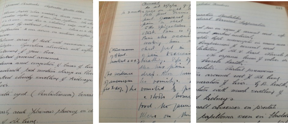
There are lots of old, stock descriptive terms that still appear in modern pathology texts – I’m thinking of things like “anchovy sauce” to describe the contents of an amoebic liver abscess.

Anchovy sauce was apparently a popular condiment in Europe and Britain from the 17th century onwards, so it would’ve been familiar to medical men of those times. Today, not so much. While anchovy sauce may still be on the shopping list for British foodies, it’s practically unknown in the rest of the English-speaking world. What we know well here in South Africa is anchovy paste (or just “fish paste”); along with peanut butter it‘s a standard sandwich filler.

But I remember clearly that as students we were lectured on the difference between anchovy sauce and anchovy paste and that only the sauce was relevant to amoebiasis. The sauce is apparently chocolate-brown and thinnish in consistency, while the paste is pinkish and has the consistency of, well, paste. But a quick browse on the web shows that today anchovy sauce and anchovy paste are employed almost interchangeably in relation to amoebic liver abscess, and even the descriptions of the appearance of sauce and paste seem to overlap. This suggests that many people have no experience of one or the other.

The anchovy sauce description makes the distinction between amoebic “pus” and the thicker greenish-yellow pus found in a bacterial liver abscess. In fact, some esteemed textbooks don’t even mention the classic anchovy sauce appearance; for example in “Pathology of Tropical and Extraordinary Diseases”, amoebic liver abscesses are said to contain “yellow or gray, opaque liquid material”. To add to the confusion, anchovy sauce is also sometimes used to describe the blood-and-pus liquid stools in amoebic dysentery (and even other types of colitis).

So the question is, if anchovy sauce no longer has any helpful resonance with the vast majority of medical students or even pathologists, why do we keep referring to it? Should we not just describe the pus in an amoebic liver abscess as reddish-brown and thinnish in consistency? (Skip the chocolate since it could be milk or dark…)

millet grains 
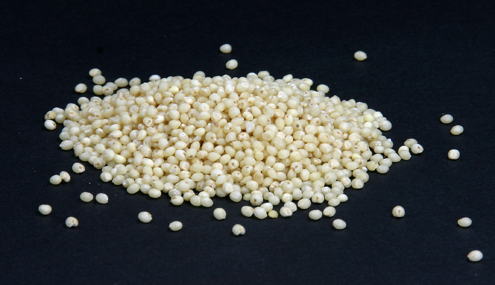
Continuing the theme but changing the topic, it could be time to reconsider the term “miliary” tuberculosis. Miliary tuberculosis occurs when a shower of blood-borne bacteria result in numerous tiny, white tuberculous foci in the organs, evidently resembling millet seeds. Now millet is not your run-of-the-mill grain these days, though students from rural Africa or India would be acquainted with it. For urban youth a grain like quinoa is more on trend, or perhaps sesame seed is more widely known, and both are much the same thing as millet visually. But the point is, when you have to explain what millet is, surely it’s no longer useful as an analogy.

nutmegdetail2 nutmegdetail1
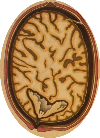
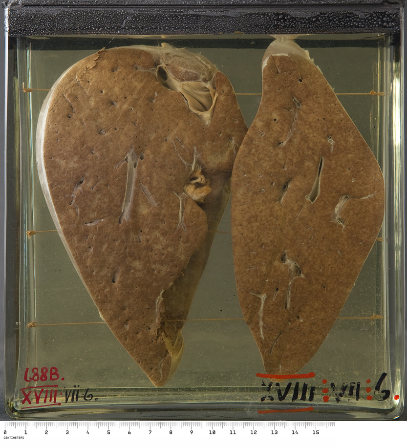
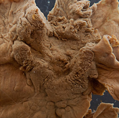
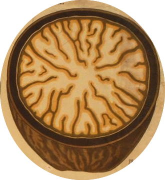
Moving on to “nutmeg liver”, a mottled appearance the liver may have when it is chronically congested, for example in heart failure. Because nutmeg was used “medicinally” through the centuries, physicians would once have known its appearance as well as their cooks did. Only someone who grates their own nutmeg will understand the nutmeg liver analogy because it refers to the appearance of a whole nutmeg in cross section. The description is no longer helpful in a modern urban world where nutmeg will almost always be bought in a convenient powdered state, ready for sprinkling.

By chance, all 3 traditional descriptive terms I’ve covered here relate to food, which is a reflection of how many of these food analogies there are. In his short article “Food in Pathology” Dr Chhanda Bewtra compiled a list of 57 examples. Some extras can be found in The Gourmet Pathologist. But questions have been raised about keeping alive some of these terms, given their lack of meaning in different cultures (See Analogies and Metaphors in clinical medicine).

And at least some pathologists feel strongly that food-related terms should be avoided all together! (http://cal.vet.upenn.edu/projects/pathterm2/colors/food.htm)

In summary, should we perpetuate descriptive terms in pathology that once had currency in a particular time, place or culture, but are an enigma to contemporary medical students?

Still, if you’ve got the time and inclination to figure them out, the old descriptions are quite cool.


REFERENCES

Chapman H, Connor DH, editors. Pathology of Tropical and Extraordinary Diseases. Washington: Armed Forces Institute, 1976.

Bewtra C. Food in pathology. Am J Dermatopathol 1996; 18(5): 555.

Batistatou A, Zolota V, Scopa CD. The “Gourmet” Pathologist. Int J Surg Pathol. 2000; 8(4):341-342.

Masukume G, Zumla A. Analogies and metaphors in clinical medicine. Clin Med 2012;12(1):55-6.

Dipping into old Cape autopsies Part 2

In December 1920 there is a confident new handwriting in the PM book, and the cases are initialled GBB. George Bertram Bartlett was the second appointed professor of pathology at the University of Cape Town, the first to actually take up the job. It appears he hit the ground running, performing 7 autopsies before the year was out.

Prof GB Bartlett’s copperplate

A summary of the primary findings for the 12 medical autopsies of 1920:

Infections:  Disseminated tuberculosis (two cases), bacterial endocarditis, septicaemia with multiple lung abscesses, dysentery, bacterial meningitis (secondary to an ear infection?).
Neoplasms: A kidney tumour, a pancreatic tumour, carcinoma of stomach (a gastrostomy had been performed by Professor Saint, first professor of surgery at UCT).
Other: Nephritis with pulmonary oedema, perforated gastric ulcer, pulmonary embolus (following a fractured tibia and fibula, which was plated – see original notes above).

It is not a profound observation, but just sad to think that deaths could have been avoided had there been effective antibiotics available.

Today, we still hold two preserved specimens from these twelve cases, the earliest specimens in our pathology teaching collection – a  Hydrocoele of the tunica vaginalis (an incidental finding) and a Carcinoma of the stomach .

X_III_1_crop  xv_iv_2_r

The following year (1921) autopsies rose to 58, from which 13 specimens are preserved. During the 1930s autopsy numbers ramped up to around 250 per year, remained at this level through the war years, but by 1950 exceeded 500 annually.


And other examples from the 1920's, when doctors could still write
Some other pages from the 1920’s, proving that doctors could once write

Dipping into old Cape autopsies Part 1

This is the lovely end paper of the first PM book of the Department of Pathology, University of Cape Town. A passing resemblance to lung alveoli at low power?
This is the lovely endpaper of the first PM book of the Department of Pathology, University of Cape Town. A passing resemblance to lung parenchyma at low power?

Along with our collection of teaching specimens, we have kept the historic post mortem books of the UCT Pathology Department – it’s amazing what medical and social history is reflected in those old reports. The first PM book opens in November 1919 and just five autopsies were recorded for that year. The youngest subject was a 10 month old boy who died of bronchopneumonia, the oldest an 81 year old man who died of infection complicating gall stones. The three other victims succumbed to bacillary dysentery, amoebic dysentery and cirrhosis of the liver.

When reading through the notes, I wondered why laboratory investigation for these cases was limited to identifying micro-organisms (such as “dystenteric organisms of the shiga type”), with no microscopic examination of the tissues i.e. histopathology. The autopsies are signed off by AS Strachan, and it turns out Dr Strachan was a bacteriologist. He must have been put in the position of conducting autopsies because the first appointed Professor of Pathology at the new medical school had sadly died, just after arriving in Cape Town in October 19181. Dr WB Martin was lost to the 1918 ‘flu, along with 400 other souls per day in Cape Town while the epidemic raged in October/ November of 19182.

This cartoon appeared in the newspaper Die Burger on 16 October 1918, and depicts the “Spanish ‘flu” as the grim reaper, Table Mountain in the background.

The Pathology department had opened in 1918, along with the departments of Bacteriology and Pharmacology, to provide the third year courses for the degree in medicine (modelled on British medical schools)1. After Dr Martin’s death there was a long delay in finding another professor for Pathology, apparently due to a scarcity of pathologists3.  Dr Strachan soldiered on as acting prosector, writing up five more medical autopsies in 1920. He also briefly noted some cases of unnatural death that passed though the morgue – two drownings on the same day, and one case of Lysol poisoning. Lysol is a disinfectant solution; according to Wikipedia it was advertised as an effective countermeasure to the influenza virus during the 1918 ‘flu pandemic. Unfortunately it was also a well known means of suicide (The Melbourne Argus 10 Jan 1912).

To be continued…

References

  1. Louw JH. In the shadow of Table Mountain. A history of the University of Cape Town Medical School. Struik: Cape Town, 1969.
  2. Phillips H. Plague pox and pandemics. Johannesburg: Jacana, 2012.
  3. Passing Events. South African Medical Record, 14 August 1920 p.296

Person in a bottle

When they first encounter our collection, I think a lot of visitors wonder, at least transiently, about who these people were. I guess this reaction is most likely when looking at the bits we associate strongly with the self, like a heart or an intact brain, and less likely when viewing, say, a pancreas or gall bladder.

For the sake of confidentiality, our specimens are anonymised and any obvious identifiers are removed from their history. Still, a few personal details may slip into the records, giving just a hint about the life and more often the death, of the person in the bottle. This is what a UCT fine arts student, Juliet Forsyth, was looking for when she compiled “Exposing individuality”.

“I went searching for shreds of humanity that were captured amongst the clinical data which I then linked to the specific specimen kept at the Centre.”

Such detail turned out to be incredibly scarce, and a hunt through the records of approximately 3500 catalogued and uncatalogued specimens in the general pathology section turned up only 10 that Juliet felt were informative. “I attached (the) sentences which hint at the personality or life of the individual to the organ that remains, so that their life may be remembered when looking at these very detached objects.”

She transposed the text onto the specimen bottles, and one comes across these particular bottles unexpectedly among all the others. Some examples are shown in the slides below:

Another artist working in the genre of “art meets science”, Karen Ingram, has had a similar instinct with regards to the Hunterian museum. Hunter seems to have documented quite a bit about his patients, and since they died over 200 years ago, the need for confidentiality has arguably lapsed.

Ingram’s short film “narrative remains” translates the records into imagined commentary from the patients themselves, so reviving what is known of their histories.

Of hibernation and hibernomas

XXIII_iii_19_newR_medium
A hibernoma found incidentally behind the left kidney of a 42 year old man, closely associated with the phaeochromocytoma which was the reason for surgery.

I’d never heard of a hibernoma until I came across this specimen in our collection. The distinct dark tan nodule at top is a phaeochromocytoma, but the flabby grey lobulated mass that makes up the bulk of the specimen is a hibernoma.

And yes, hibernoma derives from hibernation.

A hibernoma is a benign tumour composed of the same heat-generating fat or brown adipose tissue (BAT) found abundantly in hibernating mammals. Since we are mammals, it’s not too surprising that humans have (BAT). It’s well known that BAT is important in newborns (who are very susceptible to cold), but at one time it was thought that BAT disappeared by adulthood. But brown fat depots in adults can be visualised on positron emission tomography (PET) scans in the same areas as it occurs in neonates, and especially if the person was in a chilly environment when the scan was done!

Hibernation is a state of inactivity and metabolic depression in endotherms.  Hibernation (n.)1660s, from Latin hibernationem “the action of passing the winter”. [Image credit: Alex Martin]
Sites of brown adipose tissue in adult humans, as revealed by PET scanning. [Image credit: Professor Jan Nedergaard, The Wenner-Gren Institute,The Arrhenius Labs, Stockholm University]
Brown vs. White, and Brite:      

BAT appears brown due to the iron content of its many mitochondria. At a microscopic level brown fat differs from white fat in appearance, and unlike white fat has a rich vascular and nerve supply. Brown and white fat have a different histogenesis or cellular origin (see the origins of BAT). The latest finding is that adults have classical or constitutive BAT as well as an inducible “brite” (brown in white) form of BAT interspersed in ordinary white fat.

Microscopy of a hibernoma – the name coined by Louis Gery of the institute of anatomy at Strasbourg in a 1914 case discussion. Image credit: Nephron
Microscopy of a hibernoma showing the multivacuolated brown fat cells. The name “hibernoma” was coined by Louis Gery of the Institute of Anatomy at Strasbourg in 1914. [Image credit: Nephron]
Phaeochromocytomas and hibernomas are both fairly rare, and their co-occurrence in a significant number of cases is not co-incidence. Phaeochromocytomas are tumours of adrenal gland neuroendocrine cells, usually producing high levels of nor-adrenaline, and nor-adrenaline stimulates thermogenesis. It seems probable that extreme and chronic thermogenic stimulus from a phaeochromocytoma leads to BAT cellular proliferation, giving rise to a hibernoma, often adjacent to the phaeo. In support of this theory is the confirmation that the BAT in hibernomas is metabolically active.

This is all very interesting, but currently the real excitement is around BAT being potentially manipulable for the control of diabetes, obesity and the metabolic syndrome. Weight loss is a prominent symptom in patients with phaeochromocytoma, and the stimulation of BAT may account for a good deal of it. BAT consumes more glucose than any other tissue and even small amounts substantially raise the metabolic rate. BAT can be switched off by a beta-adrenergic blocker (the sort commonly used to lower blood pressure).

I’ve studiously avoided the biochemistry of thermogenesis in BAT, which underlies this whole discussion, but it hinges on the unique uncoupling protein 1 (UCP1), also known as thermogenin, which operates in BAT mitochondria to divert the production of cellular energy (ATP) into the release of heat. Being able to switch on BAT is an enticing idea, and activating UCP1 is probably the key to such a designer drug.

References / readings

Nedergaard J, Bengtsson T, Cannon B. Unexpected evidence for active brown adipose tissue in adult humans. Am J Physiol Endocrinol Metab 2007; 293(2):E444-52

Osborne AB, Johnson MH. Hibernoma, a special fatty tumor; report of a case. Am J Pathol 1949; 25(3):467-79

Nedergaard J, Cannon B. The changed metabolic world with human brown adipose tissue: therapeutic visions. Cell Metab 2010; 11(4):268-72

Lean ME, James WP, Jennings G, Trayhurn P. Brown adipose tissue in patients with phaeochromocytoma. Int J Obes 1986; 10(3):219-27.

English JT, Patel SK, Flanagan MJ. Association of pheochromocytomas with brown fat tumors. Radiology 1973; 107:2 279-281.

Vijgen GH, Bouvy ND, Smidt M et al. Hibernoma with metabolic impact? BMJ Case Reports 2012 Aug 21.

Borrowed organs

For our opening post I’ve homed in on some specimens that are interesting because of their place in medical history, specifically the history of xenotransplantation.

After his exceedingly well known first human heart transplant in 1967, Dr Chris Barnard continued experimenting. The rationale for the “piggy-back heart transplant” or heterotopic cardiac transplant is clear from this excerpt:

“In 1973, Barnard performed a heart transplant and the donor heart failed to function satisfactorily, so the patient died in the operating theatre. When Barnard came out to break the sad news, he was asked why he could not put the old heart back, as at least it had kept the patient alive. This struck Barnard as a distinct possibility. If the patient’s own heart had been left in place, and the transplant was inserted as an auxiliary pump, failure of the donor heart may not have caused the patient’s demise. Furthermore, during severe rejection episodes, which were common in those early days and a major cause of the poor results at the time, the native (i.e. patient’s own) heart might be able to maintain the circulation while rejection was reversed by increased therapy.”1

One thing led to another:

“On two occasions in 1977, when a patient’s left ventricle failed acutely after routine open-heart surgery and when no human donor organ was available, Barnard transplanted an animal heart heterotopically. On the first occasion, a baboon heart was transplanted, but this failed to support the circulation sufficiently, the patient dying some six hours after transplantation. In the second patient, a chimpanzee heart successfully maintained life until irreversible rejection occurred four days later, the recipient’s native heart having failed to recover during this period. Further attempts at xenotransplantation were abandoned and even now, more than 30 years later, xenotransplantation remains an elusive holy grail despite decades of research.”1

These are those two ragged-looking but seminal xenotransplants, preserved in the UCT pathology teaching collection:

Barnard’s own report of these two cases makes fascinating reading2.

In a similar vein, and from a similar time, this liver specimen dated 1968 is from a patient who suffered severe (sub)acute liver failure and went into coma. The catalogue description reads: “The liver is seen to be markedly reduced in size (885g), with the bulk of the surviving regenerated liver present as a large mass in the right lobe with occasional smaller nodules present elsewhere; the left lobe is shrunken, and slightly congested.”

But what is notable about this case is that a baboon liver perfusion had been performed, though unfortunately without response. The objective would have been to try to tide the patient over the acute liver failure, giving their own liver a chance to regenerate enough to resume functioning – analogous to the use of transient renal dialysis in acute kidney failure.

Between 1964 and 1970, one hundred and thirteen patients who had received extracorporeal liver perfusion or ECLP were reported (this case not among them). By 2000 the number reported was 270. Pig livers were most often used, but on review, baboon or human livers gave better long term survival (≈40% vs. ≈20%). But during this period the overall survival of acute liver failure patients receiving ECLP was no better than that of patients receiving conventional intensive care (≈25% for both)3. Today, artificial and bioartificial liver support can be part of the intensive care for acute liver failure, often as a bridge to liver transplantation, the optimal treatment.  ELCP using whole human livers (not suitable for transplant) or transgenic pig livers is still an option for temporary liver support in this context, despite the technical challenges and concerns about the risk of transmission of infectious agents4.

The availability of non-human primates for medical research is now far more limited than it was in the second half of the 20th century from when these cases date, but aside from non-human primates, other animals appear to remain “fair game” in the modern field of xenotransplantation.

See also

 http://www.humanxenotransplant.org

Benatar D. Duty and the beast: animal experimentation and neglected interests. Q J Med 2000: 93:831-835

References

  1. Brink JG, Hassoulas J. The first human heart transplant and further advances in cardiac transplantation at Groote Schuur Hospital and the University of Cape Town. Cardiovasc J Afr. 2009; 20(1):31-5
  2. Barnard CN, Wolpowitz A, Losman JG. Heterotopic cardiac transplantation with a xenograft for assistance of the left heart in cardiogenic shock after cardiopulmonary bypass. S Afr Med J. 1977; 52(26):1035-8.
  3.  Pascher A, Sauer IM, Hammer C, Gerlach JC, Neuhaus P. Extracorporeal liver perfusion as hepatic assist in acute liver failure: a review of world experience. Xenotransplantation 2002: 9: 309–324 
  4. Naruse K, Nagashima H, Sakai Y, Kokudo N, Makuuchi M. Development and perspectives of perfusion treatment for liver failure. Surg Today. 2005; 35:507–517