Category Archives: Medical History

Of mushrooms and men

Of the many natural hazards in Africa, mushrooms are not necessarily the first thing to spring to mind. But Neil van der Reij, a mushroom researcher with the Department of Agriculture and Environmental Affairs, is quoted as saying mushroom poisoning is “common” in South Africa (1).

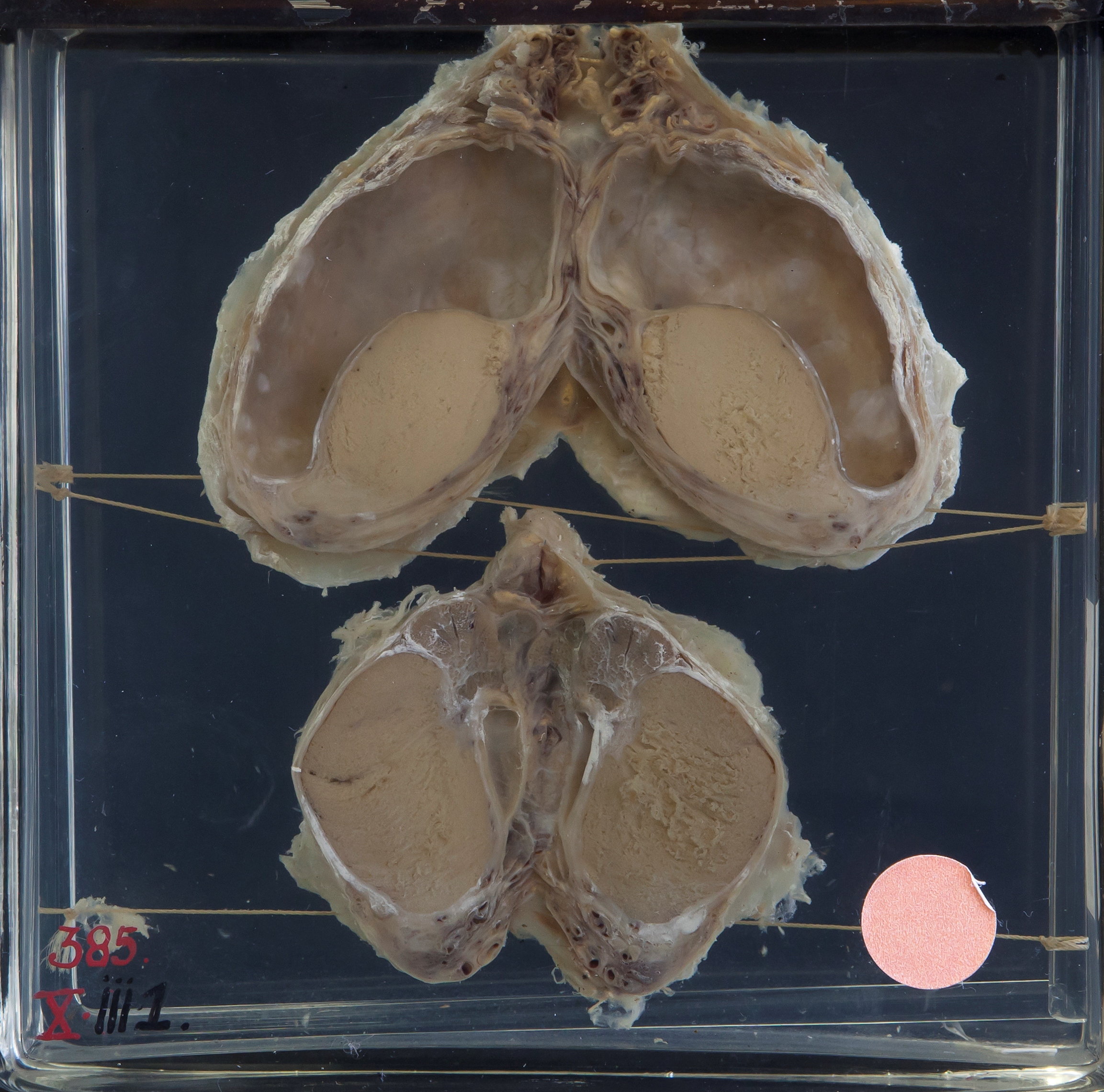
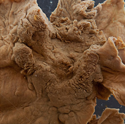
In our collection we have two liver specimens titled “mushroom poisoning”. Each case is noted to be “one of a whole family who were poisoned by eating mushrooms”. The incident occurred in Cape Town in May of 1927.

XVIII_VII_6
The preserved liver of the last of a family of 7 who died of mushroom poisoning in Cape Town in 1927.

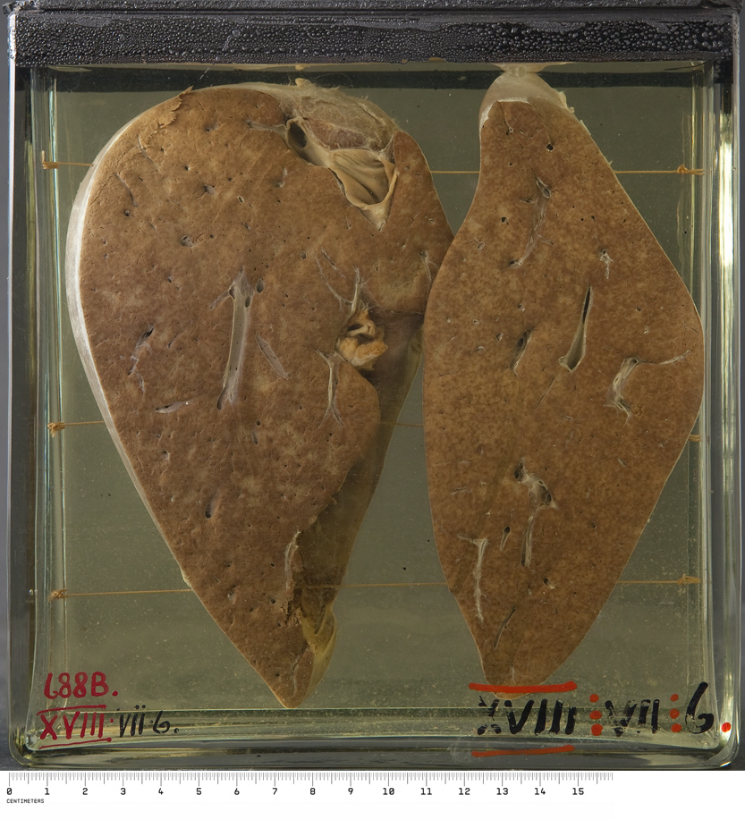
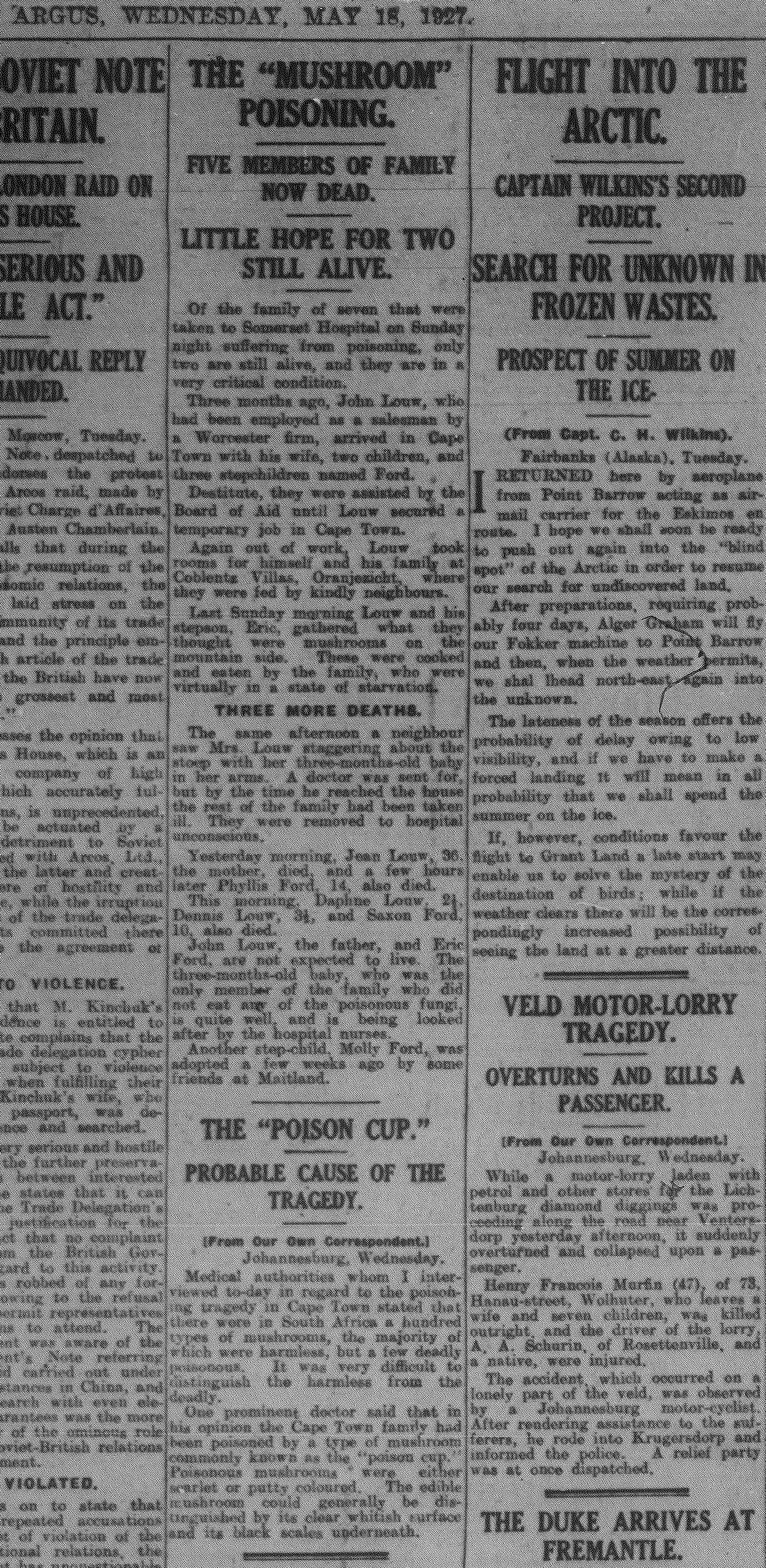
The original post-mortem reports add minimal non-pathological details, so over the Christmas period I took the time to browse the archive of the The Cape Argus for more about the story. Sure enough, a report appeared on 16 May, the day after the the entire family was admitted to Somerset Hospital. Subsequent updates documented the deaths of first the mother, followed by five children aged 2 to 14 years, then the father, and finally the 18-year-old stepson on May 19. According to the paper, the destitute family had recently arrived in Cape Town and were staying in a boarding house. Apparently they were relying on the kindness of neighbours for food. One Sunday morning the father and his elder stepson climbed the foothills of Table Mountain above the city, where they found mushrooms – it appeared these were eaten after being boiled, and symptoms set in after only a few hours. At post-mortem, the pathology was primarily fatty degeneration of the liver, kidneys and heart, resulting in multi-organ failure. The only family members to survive were the 3-month-old baby and a little girl who had been adopted by friends a few weeks before the incident.

18 May 1927 _

Of all the cases in our collection, why do these resonate particularly? Aside from being terribly sad events, there is something fascinating about natural poisoning whether by snakes, arachnids, plants or funghi. My quick search of the web found media accounts of 16 fatal cases of mushroom poisoning (in 5 clusters) in South Africa between 2008 and 2014. In addition, in 2001 a child poisoned by mushrooms was saved by a liver transplant at Red Cross Children’s Hospital, but her grandfather succumbed (2). Going back 100 years, it is part of Southern Cape history that the prominent businessman who founded the village of Wilderness died there as a consequence of eating mushrooms he had collected locally; his sister and a guest succumbed too (3). Medical reports can be found in the SAMJ archive: In 1955, five of nine related patients died in Ermelo, and in 1988, four of five patients died in the Ciskei after apparently sharing a single mushroom (4,5).

One wonders how many other cases go unrecognised.

Frustratingly, only the more recent end of the SAMJ archive is searchable, but by chance I discovered that a report of our own mushroom poisoning cases was published in November 1927 by Mervish and Silberbauer (well known Cape doctors), including the post mortem notes of Prof JB Ryrie (6). They conclude their paper with an account of experiments they conducted on a frog heart and rabbit intestine, using the vomitus of the patients.

mush book cover

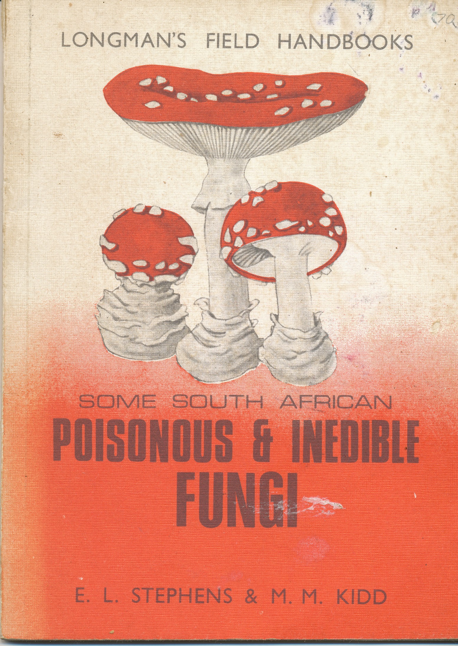
There is a pattern to these poisoning incidents; they all involve clusters of family and sometimes extend to friends as well, because a meal, with or without mushrooms, is usually shared. Also in common is that all the incidents were known or assumed to have involved Amanita phalloides, and the course to death was agonising. In the Cape, mushroom poisonings mostly occur in April/May, while in the rest of the country they occur during the months of summer rainfall. The 2016 mushroom season in Cape Town is now open for those of us who dare forage!

See also

www.pathologylearningcentre.uct.ac.za/harmful-substances-fungi

SOURCES

  1. http://www.iol.co.za/news/south-africa/kwazulu-natal/mushroom-poisoning-common-in-sa-1390479
  2. http://www.iol.co.za/news/south-africa/poisoned-girl-gets-christmas-gift-of-life-78523
  3. http://www.georgeherald.com/news/News/General/162148/Centenary-of-poison-mushroom-deaths
  4. Steyn DG, Steyn DW, Van der Westhuizen GC, Louwrens BA. Mushroom poisoning. S Afr Med J. 1956; 30:885-890
  5. Rivett MJ, Boon GP. Mushroom (Amanita phalloides) poisoning in the Ciskei. S Afr Med J. 1988;73:317.
  6. Silberbauer SF, Mervish L. Notes on cases of fungus poisoning. J Med Assoc S Afr. 1927; 1(21): 549-553

Dedicated to my colleague, friend and mushroom guide, DRH.

Dipping into old Cape autopsies Part 2

In December 1920 there is a confident new handwriting in the PM book, and the cases are initialled GBB. George Bertram Bartlett was the second appointed professor of pathology at the University of Cape Town, the first to actually take up the job. It appears he hit the ground running, performing 7 autopsies before the year was out.

Prof GB Bartlett’s copperplate

A summary of the primary findings for the 12 medical autopsies of 1920:

Infections:  Disseminated tuberculosis (two cases), bacterial endocarditis, septicaemia with multiple lung abscesses, dysentery, bacterial meningitis (secondary to an ear infection?).
Neoplasms: A kidney tumour, a pancreatic tumour, carcinoma of stomach (a gastrostomy had been performed by Professor Saint, first professor of surgery at UCT).
Other: Nephritis with pulmonary oedema, perforated gastric ulcer, pulmonary embolus (following a fractured tibia and fibula, which was plated – see original notes above).

It is not a profound observation, but just sad to think that deaths could have been avoided had there been effective antibiotics available.

Today, we still hold two preserved specimens from these twelve cases, the earliest specimens in our pathology teaching collection – a  Hydrocoele of the tunica vaginalis (an incidental finding) and a Carcinoma of the stomach .

X_III_1_crop  xv_iv_2_r

The following year (1921) autopsies rose to 58, from which 13 specimens are preserved. During the 1930s autopsy numbers ramped up to around 250 per year, remained at this level through the war years, but by 1950 exceeded 500 annually.


And other examples from the 1920's, when doctors could still write
Some other pages from the 1920’s, proving that doctors could once write

Dipping into old Cape autopsies Part 1

This is the lovely end paper of the first PM book of the Department of Pathology, University of Cape Town. A passing resemblance to lung alveoli at low power?
This is the lovely endpaper of the first PM book of the Department of Pathology, University of Cape Town. A passing resemblance to lung parenchyma at low power?

Along with our collection of teaching specimens, we have kept the historic post mortem books of the UCT Pathology Department – it’s amazing what medical and social history is reflected in those old reports. The first PM book opens in November 1919 and just five autopsies were recorded for that year. The youngest subject was a 10 month old boy who died of bronchopneumonia, the oldest an 81 year old man who died of infection complicating gall stones. The three other victims succumbed to bacillary dysentery, amoebic dysentery and cirrhosis of the liver.

When reading through the notes, I wondered why laboratory investigation for these cases was limited to identifying micro-organisms (such as “dystenteric organisms of the shiga type”), with no microscopic examination of the tissues i.e. histopathology. The autopsies are signed off by AS Strachan, and it turns out Dr Strachan was a bacteriologist. He must have been put in the position of conducting autopsies because the first appointed Professor of Pathology at the new medical school had sadly died, just after arriving in Cape Town in October 19181. Dr WB Martin was lost to the 1918 ‘flu, along with 400 other souls per day in Cape Town while the epidemic raged in October/ November of 19182.

This cartoon appeared in the newspaper Die Burger on 16 October 1918, and depicts the “Spanish ‘flu” as the grim reaper, Table Mountain in the background.

The Pathology department had opened in 1918, along with the departments of Bacteriology and Pharmacology, to provide the third year courses for the degree in medicine (modelled on British medical schools)1. After Dr Martin’s death there was a long delay in finding another professor for Pathology, apparently due to a scarcity of pathologists3.  Dr Strachan soldiered on as acting prosector, writing up five more medical autopsies in 1920. He also briefly noted some cases of unnatural death that passed though the morgue – two drownings on the same day, and one case of Lysol poisoning. Lysol is a disinfectant solution; according to Wikipedia it was advertised as an effective countermeasure to the influenza virus during the 1918 ‘flu pandemic. Unfortunately it was also a well known means of suicide (The Melbourne Argus 10 Jan 1912).

To be continued…

References

  1. Louw JH. In the shadow of Table Mountain. A history of the University of Cape Town Medical School. Struik: Cape Town, 1969.
  2. Phillips H. Plague pox and pandemics. Johannesburg: Jacana, 2012.
  3. Passing Events. South African Medical Record, 14 August 1920 p.296

Borrowed organs

For our opening post I’ve homed in on some specimens that are interesting because of their place in medical history, specifically the history of xenotransplantation.

After his exceedingly well known first human heart transplant in 1967, Dr Chris Barnard continued experimenting. The rationale for the “piggy-back heart transplant” or heterotopic cardiac transplant is clear from this excerpt:

“In 1973, Barnard performed a heart transplant and the donor heart failed to function satisfactorily, so the patient died in the operating theatre. When Barnard came out to break the sad news, he was asked why he could not put the old heart back, as at least it had kept the patient alive. This struck Barnard as a distinct possibility. If the patient’s own heart had been left in place, and the transplant was inserted as an auxiliary pump, failure of the donor heart may not have caused the patient’s demise. Furthermore, during severe rejection episodes, which were common in those early days and a major cause of the poor results at the time, the native (i.e. patient’s own) heart might be able to maintain the circulation while rejection was reversed by increased therapy.”1

One thing led to another:

“On two occasions in 1977, when a patient’s left ventricle failed acutely after routine open-heart surgery and when no human donor organ was available, Barnard transplanted an animal heart heterotopically. On the first occasion, a baboon heart was transplanted, but this failed to support the circulation sufficiently, the patient dying some six hours after transplantation. In the second patient, a chimpanzee heart successfully maintained life until irreversible rejection occurred four days later, the recipient’s native heart having failed to recover during this period. Further attempts at xenotransplantation were abandoned and even now, more than 30 years later, xenotransplantation remains an elusive holy grail despite decades of research.”1

These are those two ragged-looking but seminal xenotransplants, preserved in the UCT pathology teaching collection:

Barnard’s own report of these two cases makes fascinating reading2.

In a similar vein, and from a similar time, this liver specimen dated 1968 is from a patient who suffered severe (sub)acute liver failure and went into coma. The catalogue description reads: “The liver is seen to be markedly reduced in size (885g), with the bulk of the surviving regenerated liver present as a large mass in the right lobe with occasional smaller nodules present elsewhere; the left lobe is shrunken, and slightly congested.”

But what is notable about this case is that a baboon liver perfusion had been performed, though unfortunately without response. The objective would have been to try to tide the patient over the acute liver failure, giving their own liver a chance to regenerate enough to resume functioning – analogous to the use of transient renal dialysis in acute kidney failure.

Between 1964 and 1970, one hundred and thirteen patients who had received extracorporeal liver perfusion or ECLP were reported (this case not among them). By 2000 the number reported was 270. Pig livers were most often used, but on review, baboon or human livers gave better long term survival (≈40% vs. ≈20%). But during this period the overall survival of acute liver failure patients receiving ECLP was no better than that of patients receiving conventional intensive care (≈25% for both)3. Today, artificial and bioartificial liver support can be part of the intensive care for acute liver failure, often as a bridge to liver transplantation, the optimal treatment.  ELCP using whole human livers (not suitable for transplant) or transgenic pig livers is still an option for temporary liver support in this context, despite the technical challenges and concerns about the risk of transmission of infectious agents4.

The availability of non-human primates for medical research is now far more limited than it was in the second half of the 20th century from when these cases date, but aside from non-human primates, other animals appear to remain “fair game” in the modern field of xenotransplantation.

See also

 http://www.humanxenotransplant.org

Benatar D. Duty and the beast: animal experimentation and neglected interests. Q J Med 2000: 93:831-835

References

  1. Brink JG, Hassoulas J. The first human heart transplant and further advances in cardiac transplantation at Groote Schuur Hospital and the University of Cape Town. Cardiovasc J Afr. 2009; 20(1):31-5
  2. Barnard CN, Wolpowitz A, Losman JG. Heterotopic cardiac transplantation with a xenograft for assistance of the left heart in cardiogenic shock after cardiopulmonary bypass. S Afr Med J. 1977; 52(26):1035-8.
  3.  Pascher A, Sauer IM, Hammer C, Gerlach JC, Neuhaus P. Extracorporeal liver perfusion as hepatic assist in acute liver failure: a review of world experience. Xenotransplantation 2002: 9: 309–324 
  4. Naruse K, Nagashima H, Sakai Y, Kokudo N, Makuuchi M. Development and perspectives of perfusion treatment for liver failure. Surg Today. 2005; 35:507–517